17 Jul 2017
Caring for and managing freshly calved dairy cows – part 1
In the first of a two-part article, Nicola Gladden reviews solutions and options for dealing with postpartum problems in dairy cows.
Nicola Gladden
Job Title
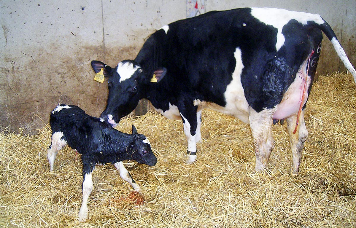
Figure 1. The calving yard/pen should be clean and dry. IMAGE: K Ellis.
Calving is a stressful and potentially dangerous event, and postpartum problems are commonly seen in dairy herds. Problems that occur in the postparturient period can adversely affect the health and welfare of the cow in the immediate term, but can also have longer-term adverse effects on production. Prompt, appropriate treatment is required to treat cows in the immediate term, but investigation into management around calving, including dry period management, is also needed to prevent future problems occurring in other animals in the herd.
This article reviews some of the evidence available regarding the care of the postpartum dairy cow and management of postpartum complications that may arise.
Postpartum problems are commonly seen in dairy cows and studies have indicated an incidence of postpartum metabolic disease approaching 40% in some herds (Ribeiro et al, 2013).
It is well recognised postpartum disease in dairy cows has an adverse effect on production (Dreyfuss et al, 1990; Duffield et al, 2009; Rutherford et al, 2016) and reduction of development of disease, combined with prompt treatment when disease does occur, is important to minimise effects on future production. It is worth remembering, with the exception of traumatic emergencies, postpartum disease is usually related to the prepartum management of the cow. When considering postpartum disease, it is important to include the farm prepartum and dry cow management as part of the investigation.
The first part of this article aims to review some of the evidence available regarding the immediate care of the postpartum dairy cow and management of postpartum complications that may arise.
Immediate postpartum management
Following parturition, the cow should be moved out of the calving pen as soon as is reasonably possible. The exact timing of movement will depend on a number of factors, including the health status of the cow and the type of fresh cow accommodation available. Measures should be undertaken, such as ensuring the cow has free access to clean water and good quality food, to try to maximise dry matter intake (DMI) following calving. This aims to mitigate the natural reduction in DMI that occurs following parturition and avoid postpartum complications that can arise as a result of negative energy balance.
It is well documented the postpartum period is a period of immunosuppression (Mallard et al, 1998) and the cow should be housed in clean, dry accommodation to minimise the risk of being exposed to infectious agents during this time (Figure 1) – this includes Johne’s disease.
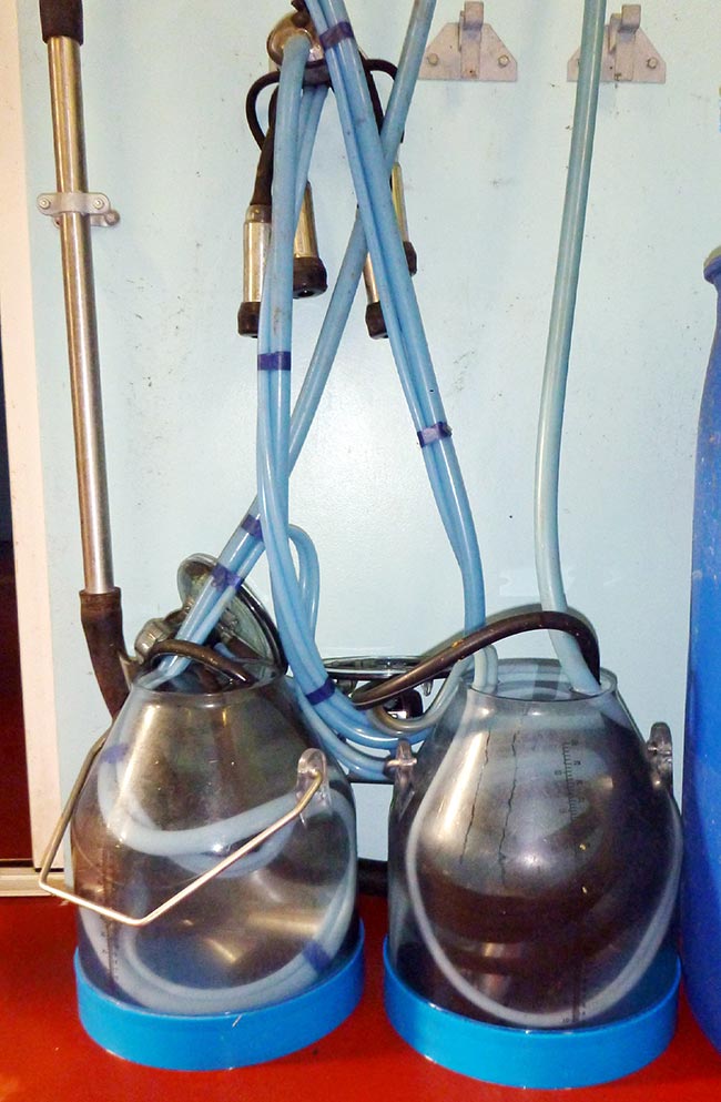
Cows determined to be at high risk to Johne’s disease should be calved separately to low-risk cows. Cows should be milked at the first opportunity post-calving and the farm should have a separate dump line for fresh cows to prevent cross-contamination of colostrum with milk from cows receiving medical treatment (such as cows with mastitis; Figure 2).
Care of the calf is outwith the scope of this article, although it is important to ensure the calf receives colostrum of adequate quality and quantity within the first four hours of life to ensure adequate passive transfer of immunoglobulins. A UK study found a prevalence of failure of passive transfer of 26% on the farms recruited (MacFarlane et al, 2015), suggesting room for improvement exists in this area on UK farms.
Analgesia
It is well recognised parturition is a painful event, although few published data exist on the use of periparturient analgesia in dairy cows, which has been highlighted in some reviews of the subject (Laven et al, 2012; Mainau and Manteca, 2011).
A study demonstrated the use of meloxicam in the immediate postpartum period was associated with an increased whole lactation yield (Carpenter et al, 2016) and these findings are supported by other studies indicating the benefits of periparturient analgesia extend beyond the initial postpartum period (Farney et al, 2013; Stilwell et al, 2014). However, other studies have not found this effect to be reproducible (Meier et al, 2014); the difference in findings may well be as a result of differences in study design.
It has been suggested the use of NSAIDs around the time of calving might increase the risk of retained fetal membranes (RFMs) due to the inhibition of prostaglandin-2α (PGF-2α), which has been shown to be important for the expulsion of fetal membranes. Studies investigating the use of flunixin meglumine in the immediate postpartum period have demonstrated an increased risk of RFMs associated with the use of NSAID analgesia (Newby et al, 2017; Waelchli et al, 1999). However, studies that have used ketoprofen or meloxicam in the periparturient period have demonstrated no increased risk of developing RFMs (Newby et al, 2014; Richards et al, 2009).
It is possible the differences reported are due to the differences in pharmacokinetics of each NSAID. Based on the evidence available, it would be prudent to avoid the use of flunixin meglumine in postpartum animals if other NSAIDs are available. However, it is worth noting evidence available is limited and no studies exist that compare different NSAIDs under the same conditions.
No NSAIDs are licensed in the UK specifically for postpartum analgesia, although a ketoprofen product is licensed for the treatment of postparturient paresis and postparturient oedema. Use of NSAIDs for postpartum analgesia in cattle is, therefore, on the cascade and appropriate (statutory) withdrawal periods should be applied.
Postpartum emergencies
Postpartum emergencies occur sporadically and can be life-threatening for the cow; prompt veterinary attention is essential for a satisfactory outcome. This section summarises how to deal with the more common postpartum emergencies that might be encountered. A more in-depth review can be found elsewhere (Miesner and Anderson, 2008; Rees, 2016).
Uterine prolapse
Uterine prolapse is a true emergency likely to be encountered at some point by any vet in mixed or farm animal practice, and has been reported to occur following up to one per cent of calvings (Gardner et al, 1990; Rees, 2016).
Uterine prolapse most commonly occurs within the first 24 hours following parturition and is known to be associated with hypocalcaemia and dystocia (Gardner et al, 1990). When early treatment is instigated, prognosis is good with reported survival rates approaching 80 per cent (Miesner and Anderson, 2008). Unlike vaginal prolapse, a cow that experiences uterine prolapse is not any more likely to experience uterine prolapse in following years (Miesner and Anderson, 2008).
Initial telephone advice to the farmer should concentrate on keeping the prolapsed uterus as clean as possible and preventing further damage. The uterus should be covered with a clean, damp sheet or towel to prevent the mucosa drying out and to prevent further contamination. The cow’s movement should be restricted to prevent trauma to the uterus and minimise the risk of rupture of the uterine artery.
Veterinary treatment of a prolapsed uterus is aimed at replacing the uterus in a manner that is as atraumatic for the cow as possible. Epidural anaesthesia is a necessity, not only to provide analgesia for the cow, but also to facilitate replacement of the uterus. Techniques for replacement of a prolapsed uterus are reviewed elsewhere (Miesner and Anderson, 2008; Rees, 2016).
Following successful replacement, oxytocin should be administered, and antibiotic and NSAID therapy should be initiated, as well as treatment of hypocalcaemia if considered necessary (Wapenaar et al, 2011). In a review of evidence-based uterine prolapse treatment, placement of a Bühner suture was a contentious treatment option, but overall there was agreement among experts in favour of placing a suture (Wapenaar et al, 2011). Placement of a Buhner suture is unlikely to prevent reprolapse of the uterus and if this does occur, the potential exists for the suture to cause substantial damage to the uterine mucosa; it is for this reason the author generally prefers not to place a Bühner suture following uterine prolapse.
Haemorrhage
Life-threatening haemorrhage can occur following parturition in the cow. Telephone advice for first aid should aim to reduce the severity of the bleeding. If the offending vessel can be located, the farmer should be instructed to occlude the bleeding vessel by pinching it until the vet arrives (Rees, 2016).
It is more common, however, that the vessel cannot be identified and, in this situation, the farmer should be instructed to pack the vagina with a large, clean bedsheet or large towels. It is important the packing used is large enough to apply sufficient pressure to occlude the bleeding vessel and it must be made clear to the farmer how much packing is needed.
Treatment of haemorrhage is concerned with stopping the bleeding and maintaining circulating blood volume. If the bleeding vessel can be identified, artery forceps can be applied and left in place for several days until a robust clot has formed. If the vessel cannot be identified, tight packing of the vagina is an alternative treatment choice. Oxytocin can be administered to stimulate myometrial contraction and result in occlusion of smaller vessels.
Supportive care may also be necessary if a large volume of blood loss has occurred. Hypertonic saline, followed by oral fluids or IV isotonic saline, can aid in restoration of blood volume and mitigate hypovolaemic shock. In some severe cases, blood transfusion might be needed.
- This article was reviewed by Kathryn Ellis, BVNS, CertCHP, PhD, DipECBHM, MRCVS.
- Managing injuries and disease in freshly calved dairy cows – part 2